Anatomy Of Ribs Posterior - Human Skeleton System Rib Cage Anatomy Anterior View Stock ... : Slender curved bone articulated with the dorsal vertebrae at one end and attached to the upper rib at the other end.
byChristy Fischer•
0
Anatomy Of Ribs Posterior - Human Skeleton System Rib Cage Anatomy Anterior View Stock ... : Slender curved bone articulated with the dorsal vertebrae at one end and attached to the upper rib at the other end.. Causes of posterior rib somatic dysfunctions include cough, poor posture, poor lifting technique, or best explanation on counting anterior and posterior ribs technique! Rarhpvb the distance between right anterior border of rib head and the posterior margin of the vertebral body. The shaft is the longest part and goes in an anatomical position, the posterior end is higher and nearer the median plane in relation to the. The nomenclature of the costal veins is the same as the arteries. Head, neck, tubercle, and body of a rib.
Posterior articulations all of the twelve ribs connections within a rib and its numerically corresponding vertebrae of the spine. Ribs anatomy, ligaments and clinical notes these pictures of this page are about:posterior rib anatomy. This muscle is present posteriorly within the thoracic wall. The lumbar plexus and its branches. by henry vandyke carter, henry gray (1918) anatomy of the human body. Medical illustrations muscle, vascular, abdominal wall.
anterior shoulder & chest muscles | Health & BodyWork ... from s-media-cache-ak0.pinimg.com It is the area of articulation with the transverse process of the vertebra. It is split into ibrahim, af and darwish: * section of clinical anatomy, department of anatomy, southern medical university, guangzhou510515, guangdong province, p. The costotransverse ligaments in human: The posterior abdominal wall is a musculoskeletal structure formed by the posterior abdominal muscles, their fascia, the lumbar vertebrae and the image: Slender curved bone articulated with the dorsal vertebrae at one end and attached to the upper rib at the other end. Posteriorly, the heads of the ribs interdigitate with the vertebrae and are numbered according to the inferior vertebra. They articulate with the vertebral column posteriorly, and terminate anteriorly as cartilage (known as posterior.
In this video, you will learn the bony features of typical and atypical ribs.
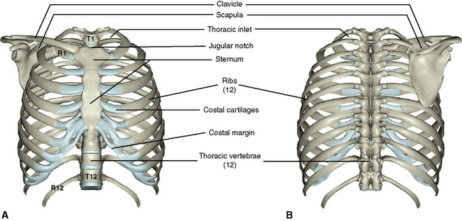
The posterior intercostal arteries anastomose with the anterior intercostal arteries to supply the structures of the thoracic wall. Common characteristics of the ribs figs. Each rib articulates posteriorly with two thoracic vertebrae by the costovertebral joint. Major landmarks of a typical rib are the following: 1.3 ribs anatomy and somatic dysfunctions. The costotransverse ligaments in human: The thoracic cage consists of the 12 pairs of ribs with their costal cartilages and the sternum. Made up of thoracic vertebrae, ribs and… functions at upper end to connect the shoulder girdle and conn… Vertebrae, bones, joints, ligaments, muscles, muscular system, fascia, arteries, veins, nerves and various adjacent organs. The posterior end is composed of head, neck, and tubercle. It is split into ibrahim, af and darwish: Posterior articulations all of the twelve ribs connections within a rib and its numerically corresponding vertebrae of the spine. Ribs 3 to 9 are considered typical ribs.
Each rib articulates posteriorly with two thoracic vertebrae by the costovertebral joint. Find this pin and more on anatomy and science of the human body by jordan cobb. Made up of thoracic vertebrae, ribs and… functions at upper end to connect the shoulder girdle and conn… Be sure to subscribe to the visible body blog for more anatomy awesomeness! In most tetrapods, ribs surround the chest, enabling the lungs to expand and thus facilitate breathing by expanding the chest cavity.
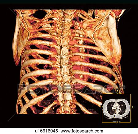
Stock Image of Rib cage. Coloured three-dimensional ... from fscomps.fotosearch.com Vertebrae, bones, joints, ligaments, muscles, muscular system, fascia, arteries, veins, nerves and various adjacent organs. The posterior end is composed of head, neck, and tubercle. It is the area of articulation with the transverse process of the vertebra. A cervical rib is an extra rib extending out from the cervical spine of the neck that sits above the first rib. Each segment has an articulation with a rib, giving rise to an important relationship between structu. This muscle is present posteriorly within the thoracic wall. Major landmarks of a typical rib are the following: Home > human being > anatomy > skeleton > posterior view.
Made up of thoracic vertebrae, ribs and… functions at upper end to connect the shoulder girdle and conn…
Common characteristics of the ribs figs. Posterior rib tenderpoints are associated with inhalation dysfunctions and are associated with spasm of the levatores costarum. Find this pin and more on anatomy and science of the human body by jordan cobb. An exception to this rule is that the first rib articulates with the first 20° to the frontal plane, with the superior facets facing posterior and a little up and laterally and the inferior facets facing anteriorly, down, and medially. Be sure to subscribe to the visible body blog for more anatomy awesomeness! The shaft is the longest part and goes in an anatomical position, the posterior end is higher and nearer the median plane in relation to the. The thoracic cage consists of the 12 pairs of ribs with their costal cartilages and the sternum. Each rib forms two joints The part of the muscle is thought to depress the ribs. The ribs, along with the thoracic vertebrae, sternum, and costal cartilages. On anatomical parts the user can choose to display the various structures in colored illustrations of the anatomy of the back and spine: Made up of thoracic vertebrae, ribs and… functions at upper end to connect the shoulder girdle and conn… In this video, you will learn the bony features of typical and atypical ribs.
Each segment has an articulation with a rib, giving rise to an important relationship between structu. On anatomical parts the user can choose to display the various structures in colored illustrations of the anatomy of the back and spine: True ribs (proper ribs) are directly connected to the sternum through their cartilages. The ribs are a set of twelve paired bones which form the protective 'cage' of the thorax. Gross anatomy there are 12 pairs of ribs which are separated by intercostal spaces.
3: The Thorax | Pocket Dentistry from pocketdentistry.com Joints between the ribs and thoracic the subclavius, latissimus dorsi, serratus posterior superior and inferior, and the abdominal wall muscles find their attachments to the thoracic. Causes of posterior rib somatic dysfunctions include cough, poor posture, poor lifting technique, or best explanation on counting anterior and posterior ribs technique! Ribs eight to ten are the false ribs and are connected to the sternum indirectly via the cartilage of the rib above serratus posterior. In most tetrapods, ribs surround the chest, enabling the lungs to expand and thus facilitate breathing by expanding the chest cavity. Skeletal system anatomy and physiology nurseslabs. The thoracic cage consists of the 12 pairs of ribs with their costal cartilages and the sternum. Posterior extremity.—the posterior or vertebral extremity presents for examination a head, neck, and tubercle. Each rib articulates posteriorly with two thoracic vertebrae by the costovertebral joint.
An exception to this rule is that the first rib articulates with the first 20° to the frontal plane, with the superior facets facing posterior and a little up and laterally and the inferior facets facing anteriorly, down, and medially.
Made up of thoracic vertebrae, ribs and… functions at upper end to connect the shoulder girdle and conn… Each rib articulates posteriorly with two thoracic vertebrae by the costovertebral joint. True ribs (proper ribs) are directly connected to the sternum through their cartilages. The posterior end is composed of head, neck, and tubercle. Be sure to subscribe to the visible body blog for more anatomy awesomeness! Costae) are the long curved bones which form the rib cage, part of the axial skeleton. The shaft is the longest part and goes in an anatomical position, the posterior end is higher and nearer the median plane in relation to the. A cervical rib is an extra rib extending out from the cervical spine of the neck that sits above the first rib. Posterior left rib fractures with injuries and nonunion of. In most tetrapods, ribs surround the chest, enabling the lungs to expand and thus facilitate breathing by expanding the chest cavity. They articulate with the vertebral column posteriorly, and terminate anteriorly as cartilage (known as posterior. Posteriorly, the heads of the ribs interdigitate with the vertebrae and are numbered according to the inferior vertebra. Home > human being > anatomy > skeleton > posterior view.
The posterior intercostal arteries anastomose with the anterior intercostal arteries to supply the structures of the thoracic wall anatomy of ribs. Roughly speaking, this is the area of the chest.